Post by petea on May 1, 2021 18:04:13 GMT
It is a surprisingly quick process, Paul, but then I have been doing it for a very long time. I think the flower image took about 15 minutes from picking it to exporting the JPEG.
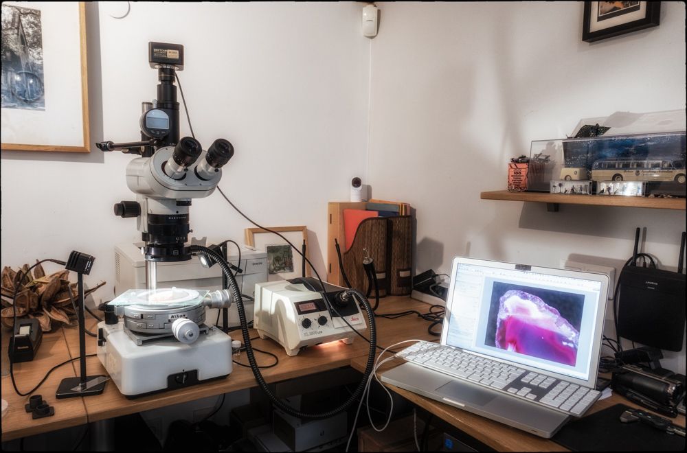
The first step is to get some light onto your subject and usually that is either a ring-light attached to the main body of the macroscope or a light positioned to either side. Normally one knows roughly where the focus height is, but if not then putting your hand under and moving it up and down until you can estimate the focal distance is a good start. Then, with the focus rack in the centre position (and ensuring the safety collar that stops the head crashing down is tightened) you can release the head and move it so that you can obtain focus with the main focus control. Now it is just a matter of orienting the specimen and lighting while looking through the eyepieces until you can either see what you need to illustrate or are happy with the appearance.
The camera in use here attaches to a computer using a USB lead and so that needs to be connected. You then open the imaging software (in this case Swift Imaging 3.0) and click on the camera in use and you will get an image on screen (actually a video stream). You can then adjust the framing by moving the specimen and focus and exposure (by either altering the lighting or changing the exposure interval) and white balance and then simply click the 'Snap' button on screen (or record video).
This will give you a single image at the point of focus set. Of course when you look through a microscope you can focus up and down continuously to help you see the specimen and this will not be reflected in a single image. However, what you can do is take a series of images at different points of focus and combine them in software to achieve a composite image with a greater depth of focus than any single frame gives. The best way to do this is the decide what is the lowest and highest points of focus and then take a series of images in between at even steps (eg maybe a 1/4 turn of the fine focus control - there are no division on the M420 whereas on many true microscopes there are). You then save all of the images in sequence and use software (it can be done manually in photoshop, but specialised software is better as it can calculate offsets etc) to create an image stack: I use either Helicon Focus or Zyrene Stacker. You can choose various ways to render the composited image and need to chose the one that is most suited to the specimen (its orientation and form will dictate that to a certain extent). After that it is a matter of saving the composite and then making any adjustments etc in an imaging programme: I usually use Lightroom.
Does that make sense / help?
There are some examples here:
www.realphotographersforum.com/forum/threads/technical-imaging-zeiss-tessovar.4309/#post-38913
www.realphotographersforum.com/forum/threads/stackshot.21091/#post-163303


